Two routes to the same cochlea
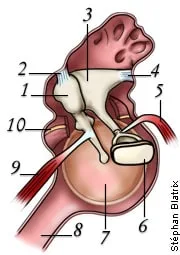
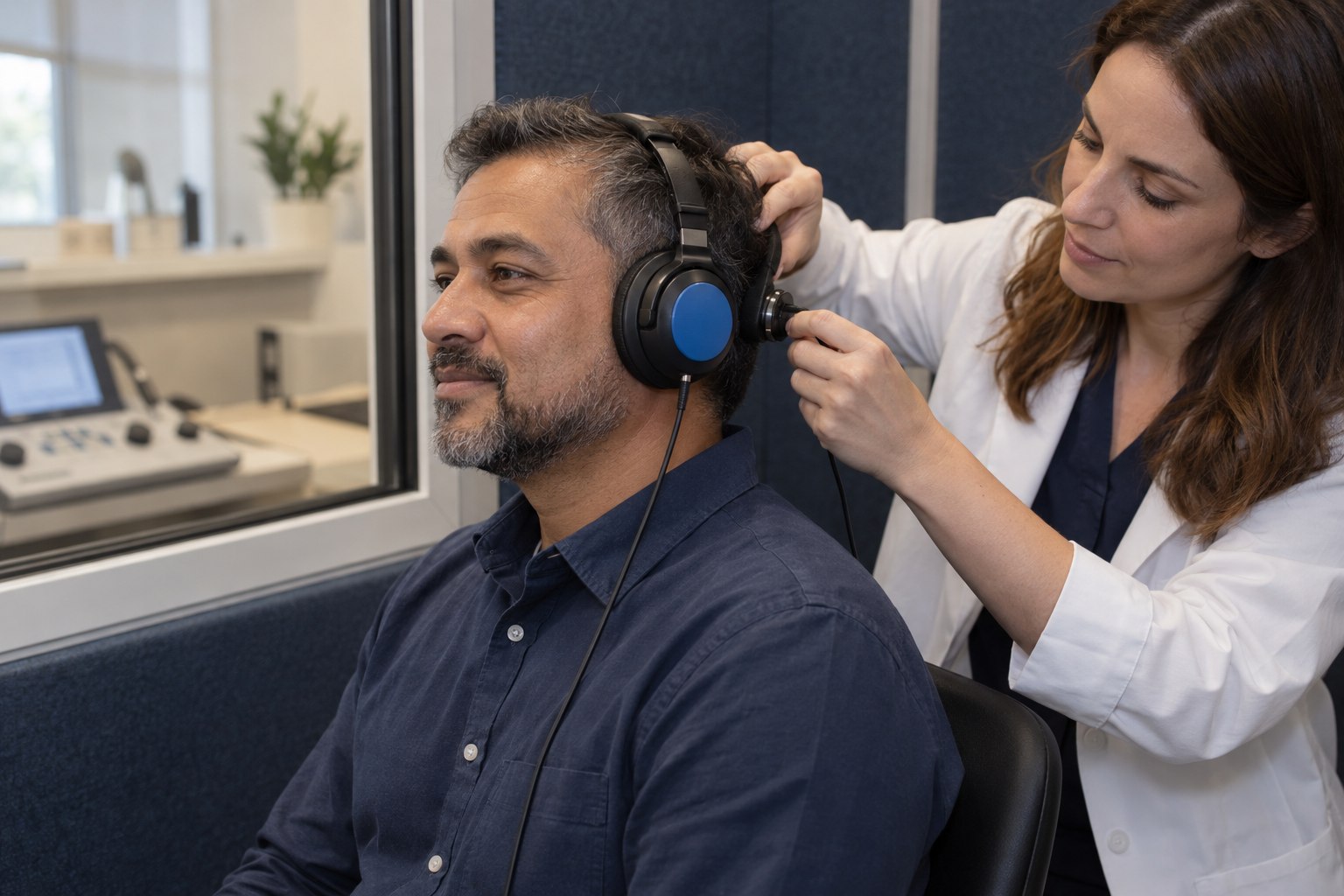
During air-conduction testing, tones travel through insert earphones or headphones, the ear canal, eardrum, middle-ear bones and inner ear. During bone-conduction testing, a small oscillator rests behind the ear or on the forehead and vibrates the skull. That route largely bypasses the outer and middle ear, but it still depends on the cochlea, auditory nerve and the person’s response.
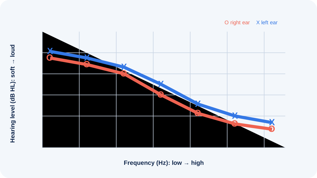
The softest level heard at each frequency is plotted in decibels hearing level. Air and bone symbols must be read by ear, frequency and masking condition. One isolated symbol is not a diagnosis.
How an air–bone gap is used
An air–bone gap is present when an air threshold is poorer than the corresponding bone threshold by more than the clinic’s expected test–retest range. It suggests that part of the reduction occurs before sound reaches the cochlea. Earwax, middle-ear fluid, eardrum changes and ossicular problems are examples a clinician may consider, but the audiogram alone cannot identify which explanation applies.
If air and bone thresholds are both reduced without a meaningful gap, the pattern can support a sensorineural loss. When both are reduced and a gap remains, the pattern may be described as mixed. Terminology should be connected to the complete evaluation.
Why masking changes confidence
Bone vibration can stimulate both cochleae. Even air-conduction sound can cross to the other ear at sufficiently high levels. Masking introduces controlled noise to the non-test ear so the clinician can be more confident that the intended ear produced the response. Reports may use brackets or other symbols to show masked thresholds.
Masking is not an optional decoration. Without it, an apparent bone threshold can represent the better cochlea rather than the poorer ear. Complex masking situations require professional judgment and sometimes retesting.
Connect the result to other evidence
Otoscopy can reveal blockage or visible ear changes. Tympanometry adds information about middle-ear mechanics and ear-canal volume. Speech measures help show how the pattern relates to communication. History identifies sudden change, drainage, pain, surgery, noise exposure or asymmetry that may alter urgency.
A person with a flat tympanogram, typical ear-canal volume and a matching air–bone gap has a coherent set of clues, but a qualified clinician still determines what follow-up is appropriate.
A worked example
Suppose right-ear air thresholds are reduced in the lower frequencies while masked bone thresholds are near the expected range. The left ear is better. The clinician confirms the symbols, checks the ear, reviews tympanometry and asks about pressure or recent illness. The useful conclusion is not “the gap proves fluid.” It is that the measured pattern supports a right conductive component that should be interpreted with the examination and history.
If repeated responses are inconsistent, the next action may simply be reinstruction and retesting.
Questions and limits
- Were the bone thresholds ear-specific and masked where needed?
- Which frequencies show a meaningful gap?
- Do tympanometry and otoscopy support the same interpretation?
- Is the difference new, asymmetric or medically concerning?
- What follow-up would change the plan?
Bone-conduction consumer products are not equivalent to diagnostic bone-conduction testing. Seek prompt care for sudden hearing change, severe pain, drainage, neurologic symptoms or marked dizziness.
How to read the printed report
Begin with the legend instead of assuming that every clinic uses identical marks. Circle, cross, bracket and arrow conventions may identify the ear, transducer, masking and a “no response” limit. An arrow does not mean there is no hearing; it means no response was obtained at the equipment or safety limit shown. Note whether inserts, headphones or a bone oscillator were used and whether reliability was rated good, fair or poor.
Next, compare air and bone at the same frequency and in the same ear. Do not infer a gap by comparing the right-ear air symbol with an unmasked bone symbol that may reflect the left cochlea. Finally, look for agreement with speech thresholds and prior tests. A new asymmetry or large change deserves explanation even when a summary average appears similar.
Prepare for follow-up
Bring prior audiograms, a medication list and a short timeline of symptoms, infections, procedures and noise exposure. Write down whether the change was sudden or gradual and whether pressure, tinnitus, dizziness, drainage or pain occurs. Ask the clinician to mark the frequencies that shaped the interpretation and explain which finding would trigger medical referral or repeat testing.
If treatment or watchful waiting is recommended, clarify the review interval and what symptoms should prompt earlier contact. Keep the original report rather than only a phone screenshot; scales, legends, transducer notes and masked symbols are part of the record. For a child or anyone whose responses were difficult to condition, the plan may include repeat behavioral measures or an age-appropriate objective test rather than forcing certainty from one incomplete session.
Sources and review notes
Sources were checked for this editorial update on October 12, 2026. External guidance can change; follow the linked source for the current version.
- ASHA — Hearing Testing
- ASHA Practice Portal — Hearing Loss in Adults
- NIDCD — Hearing, Ear Infections, and Deafness
This page provides education, not an individual diagnosis, treatment plan, legal opinion or billing decision. Historical expert contribution is credited only where supported by the original project record; it does not imply review of this update.